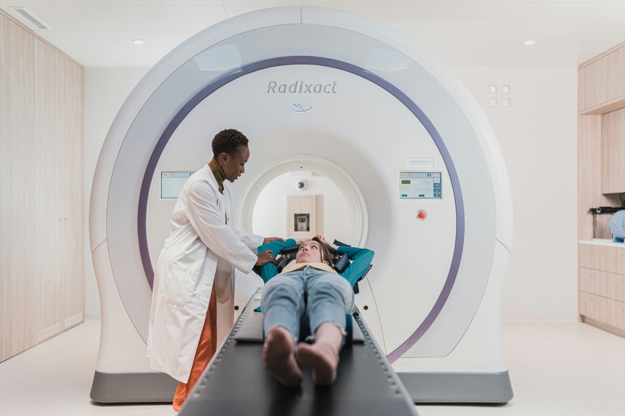
With a cancer diagnosis, imaging becomes a necessity. Imaging is how doctors determine if cancer is in remission, recurring, metastasizing or responding to treatment. It is something you will need to do on a regular basis.
In this post I will tell you about the CT scan, Pet Scan and MRI. I share important details and the pros and cons of each. Stay to the end for ways to minimize the toxic effects of these scans.
COMPUTED TOMOGRAPHY (CT)
CT scanning utilizes x-rays, a form of electromagnetic radiation. Through a complex set of computers and algorithms, the CT reconstructs cross-sectional 3-D images, or “slices” of the body, providing visualization of internal organs and structures. The thickness of the slices can be adjusted depending on what the radiologist is looking at for even more details.
Dense tissues like bone absorb more x-rays so they appear white on the resulting image. Less dense tissues like air absorb very little x-rays so they appear black. Soft tissues appear as varying shades of gray.
PROS OF CT SCANS
- Diagnosis and Staging
CT is used to identify primary tumors in various organs, including the uterus. The characteristic appearance of a tumor, including its size, shape and density can help determine if it is malignant. A CT is also beneficial to evaluate the extent of the cancer, including involvement of nearby lymph nodes or the presence of distant metastases. This helps guide treatment decisions.
- Radiation Treatment Planning
A CT scan is essential for radiation planning to ensure accurate targeting of a tumor while minimizing damage to the surrounding healthy tissues.
- Monitoring Treatment Response
A CT can assess whether a tumor is shrinking, remaining stable or progressing during and after treatment. Changes in tumor size and/or density on serial CT scans can indicate if the treatment is working or not.
- Excellent Anatomical Detail
CT excels at visualizing bone and calcifications as well as soft tissues and organs. It can provide a clear picture of the size, shape and location of a tumor.
- Speed and Availability
CTs are relatively quick to perform, which is nice. They often only take 10-15 minutes and this is very helpful for patients who may have trouble lying still for a long time. They are also widely available in most hospitals and can be used for urgent and emergency situations.
- Cost Effective
Almost every insurance company will cover the cost of CT scans for cancer patients. Compared to PET and MRI, CT scans are less expensive making them more accessible.
- Evaluation of Lung and Abdominal Structures
CT is particularly well suited for imaging the lungs, abdomen and pelvis, common sites for uterine cancer metastasis. It can pick up on tumors as small as 2-3mm in size.
- Detection of Bone Involvement
CT is highly sensitive for detecting calcifications within tumors and surrounding tissues and finding bony metastases or destruction.
- Guidance for Interventional Procedures
A CT scan can be used for minimally invasive, precise needle placement to obtain tissue samples from suspicious areas for biopsy. It can also be used for tumor ablations.
CONS OF CT SCANS
- Ionizing Radiation Exposure
The biggest drawback to a CT scan is the exposure to x-rays, which are a form of ionizing radiation. Ionizing radiation can damage DNA and lead to mutations. This increases the risk for cancer years down the road.
A millisievert (mSv) is a measure of radiation exposure. The average American is exposed to about 3 mSv of radiation from natural sources over a year. A mammogram is 0.4 mSv, which is like getting 7 weeks of exposure to natural radiation.
One CT scan of the abdomen and pelvis is approximately 31 mSv!
A single CT scan can deliver an equivalent radiation exposure to what some individuals received from the Hiroshima and Nagasaki atomic bombs! Read More
The other problem is that radiation exposure from all sources is cumulative over a lifetime and most of us are getting many CT scans so this increases our risk for developing cancer even more.
In this study in 2009 with 31,462 patients, those which had multiple CT scans >5, had an increased risk of 2.7-12% for developing cancer above the overall lifetime risk. Read More
- Limited Soft Tissue Contrast
While CT can differentiate between different soft tissues, its contrast resolution is not as high as an MRI. This can make it challenging to distinguish subtle differences between tumors and surrounding normal soft tissues in certain areas.
- Need for Iodinated Contrast Agents
In most cases, an iodine containing contrast agent is given intravenously to enhance the visibility of blood vessels, organs and some tumors. However, these contrast agents can cause allergic reactions in some people and are a risk for patients with kidney disease. There are two types, ionic and non-ionic. The non-ionic contrast is generally safer with less risk for adverse reactions.
- Limited Functional Information
While CT provides structural information, it does not assess the metabolic activity or function of different tissues. This can be a problem when it comes to differentiating scar tissue and treatment related changes from active tumors.
- Artifacts from Metal Implants
If you have any metal implants, like staples or clips in the field of imaging, this can create imaging artifacts. The artifacts appear as streaks or shadows and can obscure the nearby structures interfering with accurate interpretations.
POSITRON EMISSION TOMOGRAPHY (PET)
Unlike a CT scan, PET scans focus on the metabolic activity of cells rather than their structure. A PET scan uses small amounts of a radioactive tracer attached to a biologically active molecule, glucose, which is called fluorodeoxyglucose or FDG. The radiotracer is given intravenously about one hour before the scan.
Cancer cells have a higher metabolic rate and more glucose transporters on their surface than normal cells so they avidly take up FDG because it is a glucose analog. The scanner creates a 3D image showing the distribution of the radiotracer in the body. Areas with high radiotracer uptake correspond to increased metabolic activity, which can indicate the presence of cancer.
STANDARDIZED UPTAKE VALUE (SUV)
The SUV is a key measurement in PET scans. It’s a ratio that compares the concentration of the radioactive tracer in a specific area of your body to the concentration of the tracer in your whole body. SUV scores are not an “absolute diagnosis”. They need to be interpreted in the context of your specific cancer, medical history and other imaging results, but here are the guidelines.
- Low Uptake (SUV <2.0)
This is often considered normal background activity because many healthy tissues will have low levels of glucose metabolism. It can also indicate scar tissue after surgery or radiation.
- Moderate Uptake (SUV 2.5-5.0)
This may represent benign (non-cancerous) conditions such as inflammation or infections, which can have increased metabolic activity. It might be associated with normal physiological uptake in certain organs or the gastrointestinal tract. Although less likely, it sometimes indicates less aggressive, slow growing tumors.
- High Uptake (SUV 5.0-10)
This raises the suspicion of a malignancy as many cancers exhibit this level of increased metabolic activity. However, some inflammatory or infectious processes can also fall in this range. Further investigation may be necessary with other imaging or biopsies.
- Very High Uptake (SUV >10.0)
This level strongly suggests malignancy, often indicating a more aggressive and rapidly growing tumor with high glucose metabolism. When I had the metastatic tumor in my pelvic area prior to starting immunotherapy, the SUV uptake was 13. These areas are what are called “hot spots” and are associated with active cancer.
- SUVmax
SUVmax is the highest uptake within a specific area of interest, (tumor or lesion). An SUVmax level that is > or = to 2.5 is suspicious for a malignant tumor.
PROS OF PET SCANS
- Staging and Re-staging
PET can provide crucial information about the extent of the disease and help differentiate between localized and widespread disease.
- Detection of Recurrence
PET can help identify recurrent cancer, even in the absence of clear anatomical abnormalities on a CT or MRI scan, by detecting areas of increased metabolic activity (hot spots). In fact, a PET can often detect small metastases that may be missed by CT or MRI.
- Monitoring Treatment Response
Changes in the SUV on serial PET scans can provide an early indication of treatment efficacy before any change in tumor size may be seen on a CT or MRI. A decrease in the SUV suggests the cancer cells are becoming less metabolically active indicating a positive response.
After treatments, the PET scan can help distinguish between metabolically active residual tumor and inactive scar tissue or necrosis.
- Differentiation of Benign from Malignant Lesions
In some cases, a PET scan can help distinguish between benign lesions (inflammation, infection, scar tissue) and malignant tumors based on their metabolic activity.
- Whole Body Imaging
PET scans cover the entire body from the forehead to mid-thighs, allowing for detection of disease in unexpected locations.
CONS OF PET SCANS
- Exposure to Ionizing Radiation
PET scans involve the administration of a radioactive substance, resulting in radiation exposure. Most of the time, a PET scan will be combined with a CT scan, which exposes you to more ionizing radiation.
- Limited Anatomical Detail
The PET scan detects metabolic activity, but it can be challenging to pinpoint the exact location of abnormal metabolic activity within specific anatomical structures. This is why the PET scan is combined with a CT or MRI.
- False Positives and False Negatives
Increased metabolic activity can be seen in non-cancerous conditions such as inflammation and infection, leading to a false positive result. On the flip side, some slow-growing or less metabolically active tumors may not show significant FDG uptake, resulting in false negative results.
- Technical Factors Can Affect Results
If a patient moves during a PET scan, the image quality can be reduced. Elevated blood glucose levels can interfere with FDG uptake by cancer cells, potentially leading to false negative results. Fasting before the scan is recommended and they do check your glucose level prior to giving the injection of FDG to try to minimize this problem. The time between the tracer administration and the scan and even the type of scanner can all influence the SUV values. This is why It’s very important to have consistent scanning protocols for comparison.
- Less Availability
PET scans are more expensive than a CT or MRI and may not be as widely available. Not only that, but many times insurance won’t cover the cost of a PET scan unless it is an absolute necessity.
COMBINING PET/CT and PET/MRI SCANS
PET scan results, including the SUV scores, are always interpreted in conjunction with CT or MRI images to correlate metabolic activity with anatomical structures. This allows the radiologist to not only see areas with increased metabolic activity, but pinpoint where they are. The two scans are done at the same time or on the same day.
MAGNETIC RESONANCE IMAGING (MRI)
MRI is completely different from CT and PET scans. It utilizes a strong magnetic field and radio waves to create detailed images of the body’s internal structures. It’s a very complicated process, but an MRI gives detailed cross-sectional images of the body. Different MRI sequences can even be used to highlight specific tissue characteristics, such as water content, fat content and blood flow.
PROS OF MRI
- Excellent Soft Tissue Contrast
MRI provides better soft tissue contrast compared to CT, which allows for very detailed visualization of organs, muscles, ligaments and tumors. In fact, MRI is considered the gold standard when it comes to imaging the brain and spinal cord for the detection of metastases.
- Imaging the Pelvis
MRI is highly effective for evaluating pelvic organs, including the uterus and ovaries and rectum and can be helpful for diagnosing cancer in these areas.
- No Ionizing Radiation
MRI does not use ionizing radiation, making it a safer option for patients who require frequent imaging.
- Multiplanar Imaging
The MRI machine can acquire images in any plane without repositioning the patient, including from the front, side, top down or bottom up (like a stack of pancakes) and more. This provides a very comprehensive anatomical assessment of all the structures and organs.
- Functional Imaging Capabilities
Advanced MRI techniques can provide information about tissue cellularity and blood flow, which can be helpful in differentiating benign from malignant lesions and in assessing treatment response.
- Contrast Agent Less Toxic to Kidneys and Less Allergic Reactions
The gadolinium contrast agent used in MRI has a lower risk of damaging the kidneys than the iodinated contrast agents used in CT scans. It is also less likely to cause an allergic reaction. However, gadonlinium is not benign. More on this in the cons section.
CONS OF MRI
- Longer Scan Times
MRI scans take a lot longer than a CT. A CT typically takes 10-15 minutes, where an MRI can take up to 45 minutes depending on what is being imaged. This can be tough if you have trouble laying still for a long time.
- Claustrophobia
The MRI scanner is very enclosed and can cause anxiety if you have claustrophobia. I have this issue and have to keep my eyes closed and concentrate on my breathing during the scan to remain calm. The MRI scanner is very noisy with loud clacking and banging sounds too, which can make it even more nerve wracking. You can ask for ear plugs or headphones to help with this. Some people need a mild sedative before an MRI.
- Contraindicated with Metal Implants
The strong magnetic field of the MRI scanner can be a risk for patients with certain metallic implants, such as pacemakers. They will ask you questions about this prior to scheduling an MRI. You cannot wear anything with metal during the scan. If there is any metal it can cause significant artifacts that can obscure nearby tissues.
- Gadolinium Contrast Agent
Gadolinium is a heavy metal that is used intravenously as a contrast agent for MRIs. There are several reported complications with this agent. It is cumulative and trace amounts can build up in the brain and body over time leading to brain fog, fatigue and pain. Some people experience temporary side effects like dizziness, headache or nausea. Make sure to discuss all this with your oncologist before doing an MRI.
- Motion Artifacts
If you move, it can significantly affect the image quality. This is the hard part, staying perfectly still in a noisy enclosed tube for a long time!
- Less Bone Detail
MRI is not as sensitive as a CT for detecting calcifications and subtle bony abnormalities.
- Higher Cost and Limited Availability
MRI scans are more expensive than CT scans and some hospitals do not have MRI availability. Sometimes insurance will not cover the costs of an MRI.
WAYS TO REDUCE TOXICITY
- Melatonin
Take 300mg of melatonin one hour before a CT or PET scan to reduce radiation toxicity.
- Ask for a Low-Dose CT Scan
This uses less radiation than standard scans, but still gives the valuable information needed.
- Ask for a Non-Ionic Contrast Agent for CT Scans
This form of contrast has less side effects and is better tolerated.
- Try to Limit the Number of CT, PET or MRI Scans
I asked my oncologist to limit my scans to twice a year and I follow certain labs in between to check for recurrence.
- Ask if You can Forgo the Contrast Agent
I have done this before and they said I didn’t have to get the injectable contrast.
- Drink Lots of Water
This helps flush out toxins and protects your kidneys from damage. Follow fasting instructions, but beyond that, drink water before and after scans.
- Drink Hydrogen Water
Hydrogen has been shown to be an effective radio-protective agent in some studies. You can buy hydrogen water prepackaged or make your own. Read More
- Take Anti-Oxidants and Protective Supplements
Vitamins C, E, A , selenium, magnesium and more can protect your cells against DNA damage.
- Use Zeolite after Gadolinium Exposure
Zeolite helps detoxify the body of heavy metals.
- Consider an Alternative Scan Called Prenuvo
Prenuvo is a type of MRI. You have to pay for this out of pocket and it is not available everywhere, but there is no radiation exposure or contrast agent, which is nice.
But they that wait for the Lord shall renew their strength; They shall mount up with wings as eagles; They shall run, and not be weary; They shall walk, and not faint.
Isaiah 40:30

